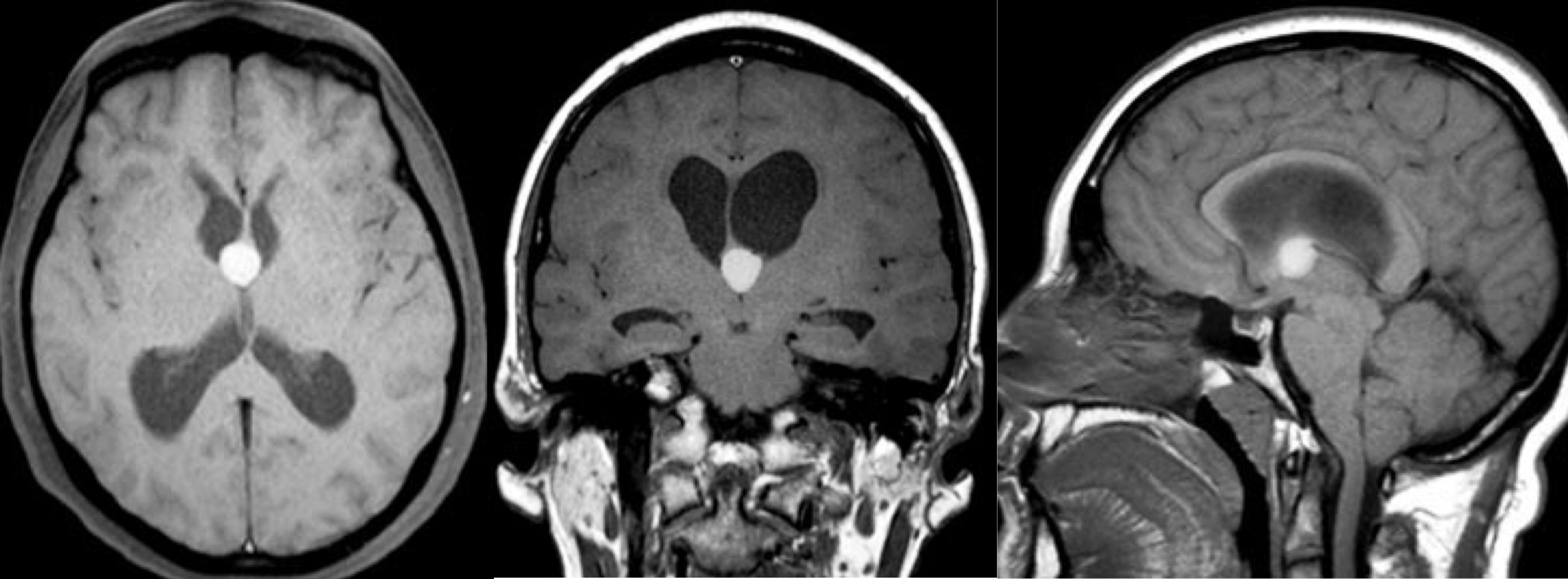
Colloid cysts are fluid-filled, rounded cystic lesions, ranging in size from a few millimetres to several centimetres for the largest, which form in the brain, usually in the third ventricle, a deep cavity of the brain in which cerebrospinal fluid circulates, making their removal a delicate task. These spherical cysts have a thin shell and are filled with a gelatinous substance called colloid (similar to glue). This content can be very fluid or relatively thick.
Colloid cysts are always benign. They are not considered tumors and are not cancerous, which means they do not spread and do not require radiotherapy or chemotherapy.
Colloid cysts, on the other hand, can increase in size over time and recur if their removal is incomplete.
Colloid cysts are very rare, affecting just 3 people per million population, and although they can be found at any age, they are usually diagnosed in adults in their 30s and 40s.
They are located in the cerebral ventricles, in which the cerebrospinal fluid circulates. In the vast majority of cases, they are located in the 3rd ventricle, opposite the orifices that connect the right and left lateral ventricles with the 3rd ventricle.
Because these cysts are located in the third ventricle, they can obstruct the flow of cerebrospinal fluid from one ventricle to the other.
They therefore most often manifest as dilatation of the lateral cerebral ventricles, upstream of the colloid cyst.
This dilation, known as hydrocephalus, usually develops very slowly.
Symptoms usually appear very gradually:
– headaches
– gait disorders
– behavioral disorders
– memory disorders
– double vision
More rarely, symptoms are much more acute and severe, in cases of sudden blockage of cerebrospinal fluid circulation. Signs of acute intracranial hypertension then develop:
– intense headaches,
– nausea, vomiting,
– drowsiness, coma.
This is sometimes an absolute emergency.
How is the diagnosis made?
Diagnosis is made on brain MRI.

Treatment
Treatment is surgical. The aim is to empty the cyst and, ideally, remove its wall to prevent recurrence. The immediate aim is to restore cerebrospinal fluid circulation.
This is a delicate surgery requiring a certain technical expertise. The cyst is removed using a microscope or an endoscope: an endoscope a few millimeters in diameter connected to a camera is inserted into the ventricles. Tools are introduced along the endoscope to gradually empty and remove the cyst.
However, most colloid cysts can be safely monitored rather than requiring surgery.
The factors that determine the choice between observation or surgical removal are symptoms, degree of ventricular dilatation, exact location, size and age.
