Radiosurgery involves highly focused, high-precision irradiation of a region of the brain presenting an abnormality (benign or malignant tumors, malformation, functional disorder), enabling neurosurgical intervention to be avoided.
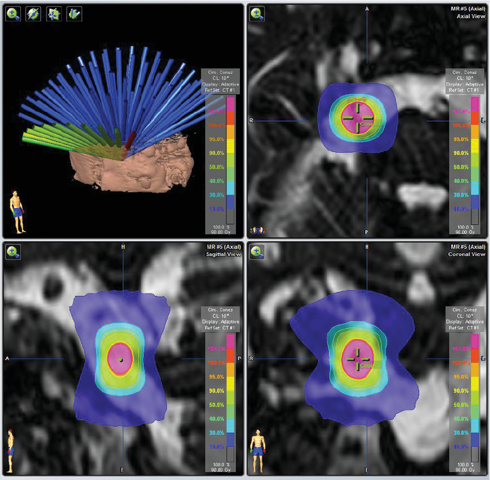
Radiosurgery is a technique for treating certain intracranial lesions, using multiple beams of very fine ionizing radiation that converge in a very precise, “surgical” way, in a single focus corresponding to the lesion to be treated. At the level of the target to be treated, where the beams are concentrated, the radiation doses delivered by the different beams add up, generating a high dose in a single session, giving the desired therapeutic effect. Surrounding and distant tissues will only be crossed by a small number of beams, and therefore only a small, non-deleterious dose of radiation.

Radiosurgery therefore has the advantage of avoiding the risks and possible complications or contraindications of conventional neurosurgical intervention for these deep lesions.
It is also an essential adjunct when surgery has not, by choice, made it possible to remove the entire lesion, in order to reduce the risks of surgery.
It is also a very useful solution in the event of residual growth or tumor recurrence, as an alternative to repeat surgery.
Radiosurgery was born of neurosurgeons’ expertise in cranial stereotaxis, which consists in locating and targeting deep intracranial zones using a three-dimensional coordinate system.
These stereotactic techniques were then applied to radiotherapy by neurosurgeons, giving rise to radiosurgery:
- Gamma-knife: the first device was invented in 1951 by a Swedish neurosurgeon (Prof. Karl Leksell).

- The Cyberknife, a “multi-purpose, multi-organ” radiosurgery device, was then developed in the late 80s by Stanford neurosurgeon Prof. John Adler.
In France, radiosurgery (LINAC) began in 1984 thanks to Prof. Betti, a neurosurgeon and student of Prof. Talairach, head of the neurosurgery department at Hôpital Sainte-Anne and a world pioneer in stereotaxy. Cooperation between Sainte-Anne Hospital and the APHP (Tenon Hospital for radiology and Lariboisière Hospital for neuroradiology) began in 1986.
Two dedicated “head and neck” radiosurgery units high-precision are currently available:
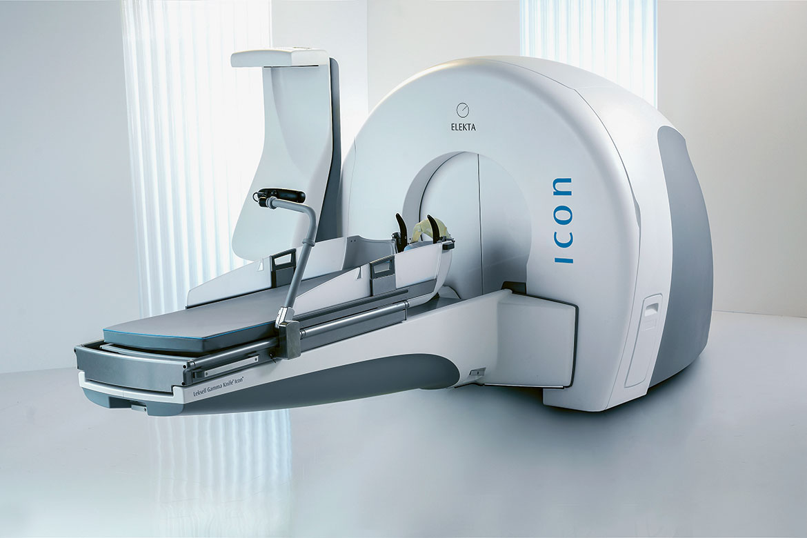
- The GammaKnife developed by Prof. Lars Leksell, a neurosurgeon at Karolinska in Stockholm. It can be used with or without an invasive frame. This technique uses range rays from a radioactive cobalt source.

- The latest-generation Zap-X, developed by Stanford neurosurgeon Prof. John Adler, uses high-energy X-rays from a linear particle gas pedal.
Other multi-purpose equipment is available for radiosurgery on other organs such as the lung, liver, prostate and spine, tailored to hospitals with highly diversified care activities.
- The CyberKnife is a radiosurgery unit also developed by Prof. John Adler, which can also be used to radiate other organs. It uses high-energy X-rays from a linear particle gas pedal.

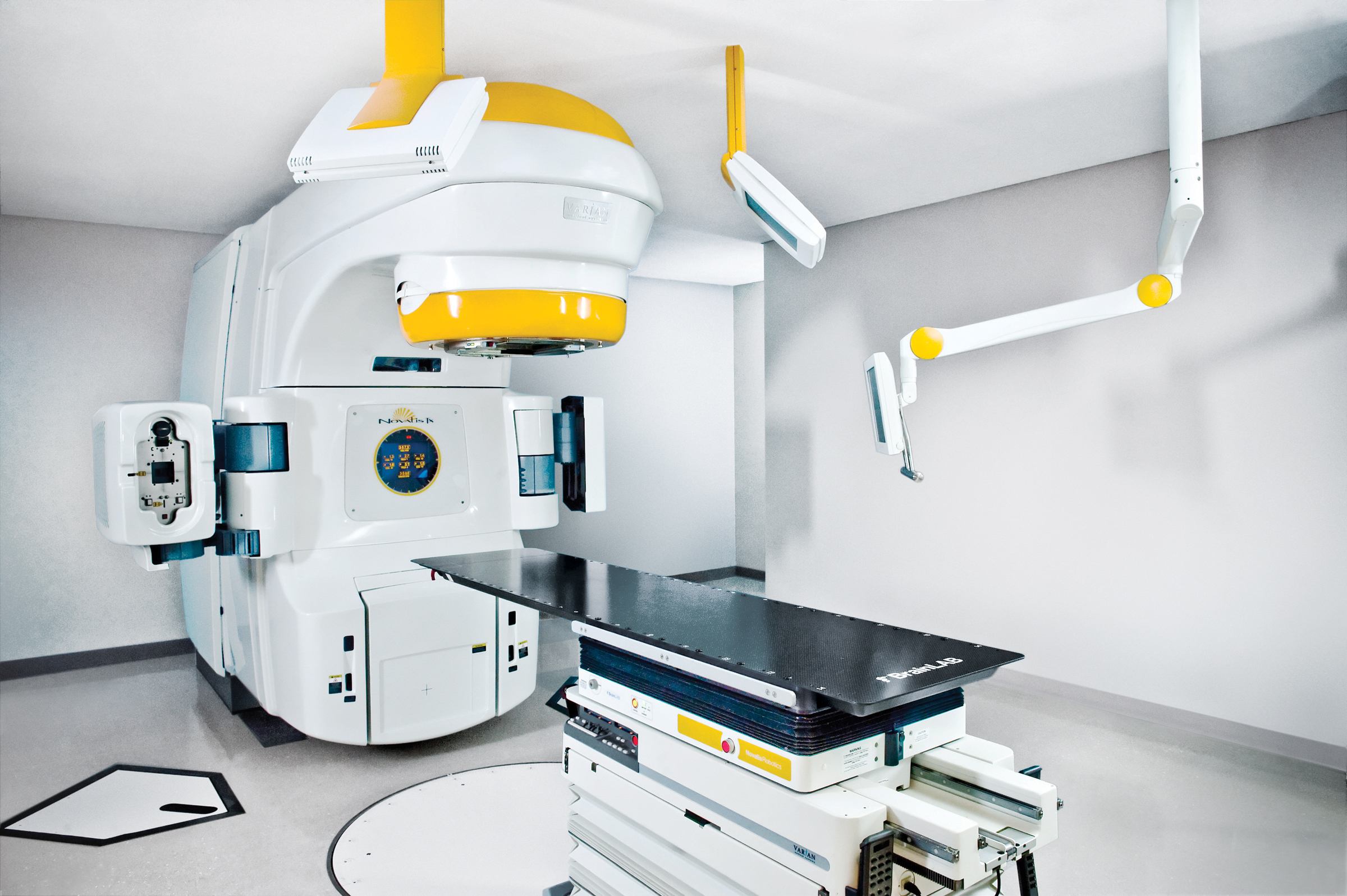
- The Novalis is a highly versatile combination of radiotherapy and radiosurgery equipment. It uses high-energy X-rays from a linear particle gas pedal.

